×
General Doppler Imaging
-
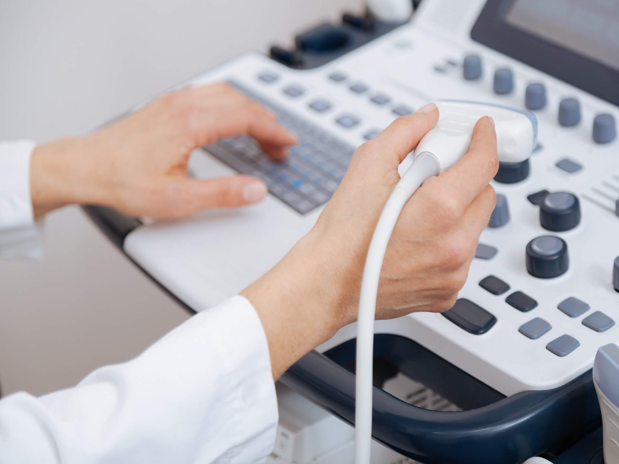
Ultrasound imaging, also called ultrasound scanning or sonography, involves the use of a small transducer (probe) and ultrasound gel to expose the body to high -frequency sound waves. Ultrasound is safe and painless, and produces pictures of the inside of the body using sound waves. Ultrasound examinations do not use Ionizing radiation (as used in x-rays). Because ultrasound images are captured in real-time, they can show the structure and movement of the body's internal organs, As well as blood flowing through blood vessels.
Conventional ultrasound displays the images in thin, flat sections of the body. Advancements in ultrasound technology include three-dimensional (3-D) ultrasound that formats the sound wave data into 3-D images. Four-dimensional (4-D) ultrasound is 3-D ultrasound in motion.
Doppler ultrasound is a special ultrasound technique that evaluates blood flow through a blood vessel, including the body's major arteries and veins in the abdomen, arms, legs and neck.
-
Why should I do it ?
-
Sonography is a useful way of evaluating the body's circulatory system. Vascular ultrasound is performed to:
- Help monitor the blood flow to organs and tissues throughout the body.
- Locate and identify blockages (stenosis) and abnormalities like plaque or emboli and help plan for their effective treatment.
- Detect blood clots (deep venous thrombosis (DVT) in the major veins of the legs or arms.
- Determine whether a patient is a good candidate for a procedure such as angioplasty.
- Evaluate the success of procedures that graft or bypass blood vessels.
- Determine if there is an enlarged artery (aneurysm.
- Evaluate varicose veins.
-
In children, ultrasound is used to:
- Aid in the placement of a needle or catheter into a vein or artery to help avoid complications such as bleeding, nerve injury or pseudo-aneurysm (abnormal outpouching of an artery with the risk of rupture) .
- Evaluate a connection between an artery and a vein which can be seen in congenital vascular malformations (arteriovenous malformations or fistula) and in dialysis fistula.
-
Doppler ultrasound images can help the physician to see and evaluate:
- blockages to blood flow (such as clots)
- narrowing of vessels
- tumors and congenital vascular malformations
- reduced or absent blood flow to various organs, such as the testes or ovary
- increased blood flow, which may be a sign of infection
-
Any preparations needed?
- You should wear comfortable, loose-fitting clothing.
- You may need to remove all clothing and jewelry in the area to be examined.
- Other preparation depends on the type of examination you will have. For some scans your doctor may instruct you not to eat or drink for as many as 12 hours before your appointment. For others you may be asked to drink up to six glasses of water two hours prior to your exam and avoid urinating so that your bladder is full when the scan begins.
- In case of children, ultrasound examinations are very sensitive to motion, and an active or crying child will slow the examination process. To ensure a smooth experience, it would be beneficial to explain the procedure to the child prior to the exam.
- If your abdominal vessels are being examined, unless the examination is performed on an urgent basis, it is best to fast before the procedure.
-
Sonography is a useful way of evaluating the body's circulatory system. Vascular ultrasound is performed to: