×
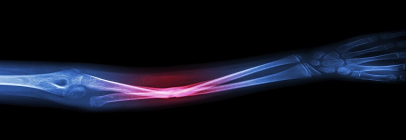
Bone X-ray

-
An x-ray (radiograph) is a noninvasive medical test that helps physicians diagnose and treat medical conditions. Imaging with x-rays involves exposing a part of the Body to a small dose of ionizing radiation to produce pictures of the inside of the body. X-rays are the oldest and most frequently used form of medical imaging.
-
A bone x-ray is used to:
- Diagnose fractured bones or joint dislocation.
- Demonstrate proper alignment and stabilization of bony fragments following treatment of a fracture.
- Guide orthopedic surgery, such as spine repair/fusion, joint replacement and fracture reductions.
- Look for injury, infection, arthritis, abnormal bone growths and bony changes seen in metabolic conditions.
- Assist in the detection and diagnosis of bone cancer.
- Locate foreign objects in soft tissues around or in bones.
-
Any preparations needed?
- Most x-ray examinations require no special preparation
- You may be asked to remove some or all of your clothes during the exam.
- You may also be asked to remove jewelry, removable dental appliances, eye glasses and any metal objects or clothing that might interfere with the x-ray Images.
- Women should always inform their physician and x-ray technologist if there is any possibility that they are pregnant.